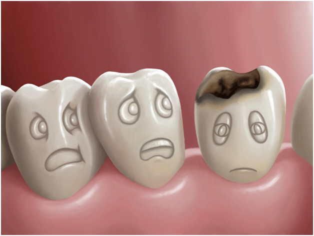

Tooth decay often begins long before a person feels pain or notices a visible hole in a tooth. In its earliest stages, enamel can begin losing minerals while the outer surface still appears mostly intact. Dentists are trained to identify these subtle changes before a traditional cavity has formed, and finding decay at this stage can make a meaningful difference: a weakened but intact tooth can often be monitored, strengthened, or treated conservatively, while a physical cavity usually requires restorative treatment such as a filling.
At Crystal Dental, early diagnosis is central to our preventive philosophy. For patients throughout Downtown Los Angeles, Koreatown, and Mid-Wilshire, the goal is not simply to find cavities. It is to recognize the earliest signs of disease so patients have more options and can preserve as much healthy tooth structure as possible.
Early Tooth Decay Starts With Mineral Loss
Before a cavity becomes visible, acids produced by plaque bacteria gradually strip minerals such as calcium and phosphate from enamel, a process called demineralization. The tooth may still be structurally intact, but the enamel becomes more porous and vulnerable. Sometimes this shows up as a chalky white area, which we cover in detail in our guide to what white spots on teeth mean. Just as often, the early lesion is invisible to the patient, and because pain develops much later, relying on symptoms alone allows the problem to progress unnoticed. The reason this stage matters is that enamel with an intact surface may still regain minerals and stabilize, which keeps remineralization-based options on the table.
Visual Examination Reveals Subtle Changes in Enamel
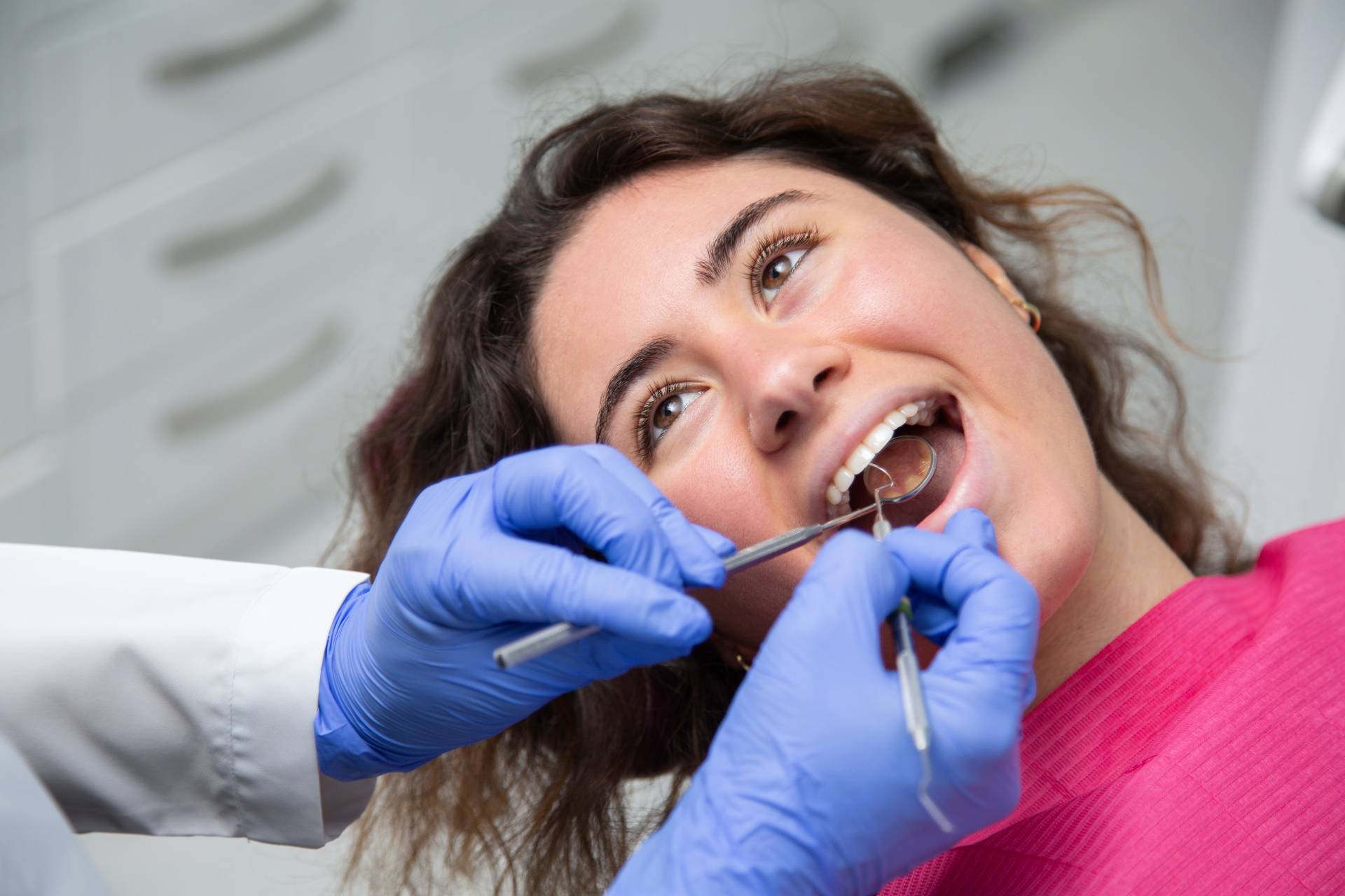
A careful visual examination remains one of the most important tools for identifying early decay. During a routine dental examination, the dentist looks for changes in color, texture, and surface appearance: early lesions may look white, dull, chalky, rougher, or less reflective than healthy enamel. The teeth are often cleaned and dried first, because demineralized enamel reflects light differently once dry, making early lesions easier to see.
Location matters as well. Areas near the gumline, between teeth, around orthodontic brackets, and within deep grooves on chewing surfaces are more vulnerable to decay. Recognizing these patterns helps the dentist judge whether a suspicious area likely represents active mineral loss or another type of enamel change.
Dental X-Rays Can Reveal Decay Hidden Between Teeth
Not all early decay can be seen directly. The spaces between neighboring teeth are especially difficult to evaluate visually because the contact points block the dentist’s view. Bitewing X-rays help here: mineral loss appears as an area that allows more radiation to pass through than healthy enamel, and the dentist can assess whether the change is confined to enamel or has progressed deeper.
X-rays are not taken on a rigid schedule. Their frequency should be based on cavity risk, age, oral health history, and clinical findings, which is the same individualized logic behind how often you should visit the dentist in the first place. A patient with frequent cavities may need imaging more often than someone with consistently low risk.
Intraoral Cameras Help Patients See What the Dentist Sees
Intraoral cameras are small devices that capture detailed images of the teeth and gums, revealing discoloration, cracks, plaque accumulation, and worn restorations that are hard to appreciate with a mirror alone.
Their biggest advantage is communication. Instead of simply being told an area needs monitoring, patients can see the exact location and appearance of the concern. That makes it easier to understand why preventive care, improved hygiene, or follow-up is being recommended, and it keeps the diagnostic process transparent, which is central to how Crystal Dental approaches patient education.
Dentists Evaluate Whether an Early Lesion Is Active or Stable
Not every area of demineralization is actively progressing. Some lesions stabilize when oral hygiene improves, dietary habits change, or mineral balance is restored. So, dentists consider more than whether a spot exists: an active lesion may look chalky or rough and sit where plaque frequently accumulates, while a stable lesion often looks smoother and shows little change over time.
This distinction drives treatment. Monitoring may be appropriate for a stable lesion, while an active one calls for focused preventive intervention based on current disease activity rather than appearance alone.
Cavity Risk Helps Guide the Diagnosis
The same enamel change can behave differently in two different patients. Someone with frequent cavities, dry mouth, high sugar intake, or reduced saliva flow is more likely to see progression, and certain medications and health conditions also raise risk. Patients in orthodontic treatment have more plaque traps, while older adults may develop decay on root surfaces exposed by gum recession.
By weighing the whole pattern of risk, the dentist can decide whether an early lesion should be monitored, treated preventively, or managed more actively. This personalized approach is more useful than treating every suspicious area the same way.
Early Detection Can Create More Conservative Treatment Options
The biggest advantage of catching decay before a cavity forms is that the enamel may still be structurally intact. The dentist may recommend improved brushing and flossing, fluoride therapy, dietary changes, or other preventive measures that reduce acid exposure and support remineralization.
For certain noncavitated enamel lesions, newer treatments such as Curodont may also be considered as part of a natural tooth healing approach. Curodont is designed to support mineral redeposition within early enamel lesions, but it is not appropriate for every patient or every area of decay, which is why professional evaluation comes first. Once a physical cavity develops and tooth structure is lost, preventive measures alone cannot replace missing enamel, and a filling or another restorative treatment may be necessary.
Early Detection Usually Costs Less Too
Conservative care for early lesions, such as monitoring, fluoride, and preventive visits, generally costs far less than the fillings, crowns, or root canals that become necessary once decay progresses, and waiting for a toothache can turn a routine finding into emergency care. Many insurance plans cover examinations, X-rays, and preventive treatment at little or no out of pocket cost, while restorative work typically involves deductibles and coinsurance.
If you are unsure what your plan covers, or you do not have dental insurance, our Payment Options page explains the insurance plans Crystal Dental accepts and the financing available, so cost does not have to delay an evaluation.
Monitoring Is an Important Part of Early Diagnosis
Not every early enamel change requires immediate treatment. In some cases the most appropriate approach is careful monitoring: the dentist documents the area with photographs, X-rays, and clinical notes, then compares it at future visits. If the lesion stays stable, more invasive treatment may never be needed. If it begins progressing, the dentist can intervene before significantly more tooth structure is affected, and well before symptoms would have revealed the problem on their own, since early decay rarely causes pain.
Monitoring is not ignoring the problem. It is an active part of conservative care, and patients play a real role in it by maintaining excellent hygiene, following dietary recommendations, and keeping scheduled examinations so changes can be tracked over time.
Frequently Asked Questions
Can dentists detect decay before it appears on an X-ray?
Yes. Some early enamel changes may be visible during a clinical examination before they are clearly detectable on dental X-rays. Dentists use visual findings, cavity risk, location, and other information together rather than relying on a single diagnostic method.
Do all white spots on teeth mean early decay?
No. White spots may result from demineralization, fluorosis, developmental enamel changes, or other causes. A professional examination is necessary to determine whether a white area represents active tooth decay.
Can early tooth decay be reversed?
Certain early enamel lesions may be strengthened through remineralization when the enamel surface remains intact. Once a physical cavity forms and tooth structure is lost, the tooth generally requires restorative treatment.
How often should early decay be monitored?
The appropriate interval depends on the lesion, the patient’s cavity risk, oral hygiene, and other clinical factors. Some patients may need more frequent evaluation than others.
Can early decay still progress even if I brush every day?
Yes. Brushing is important, but cavity risk also depends on diet, fluoride exposure, saliva flow, cleaning between the teeth, genetics, and other factors. Daily brushing should be combined with a broader preventive care plan.
Early Diagnosis Helps Preserve More Healthy Tooth Structure
Tooth decay does not begin with a hole in the tooth. It begins with subtle mineral changes that are difficult for patients to see or feel. Through careful visual examination, dental X-rays, intraoral imaging, risk assessment, and ongoing monitoring, dentists can identify these changes before significant structural damage occurs and focus on the most conservative treatment that is appropriate.
If you are concerned about early tooth decay, or it has been a while since your last examination, schedule a visit with Crystal Dental at our Figueroa Street or 5th Street office, serving patients across Downtown Los Angeles, Koreatown, and Mid-Wilshire. A careful evaluation can identify problems early, explain your options clearly, and support long term protection of your natural teeth.